Root Canal Treatment
Accurate and professional root canal treatment at HanzaDent! Root canal treatment helps save a damaged tooth and restore its health. Find out more and book an appointment at our clinics today!
Book a visitModern Technology
Best Dentists
Quick and Convenient Booking
Root Canal Treatment in Tallinn
Root canal treatment in Tallinn can be performed at three HanzaDent Dental Clinics: Tammsaare road clinic, Paldiski road clinic, and Laulupeo street clinic. Endodontic treatment is necessary when a tooth hurts or when there is an infection under the root of the tooth. In both cases, it is a complication of advanced tooth decay. Often, when there is an infection under the tooth, the patient may not experience any symptoms at all. In such cases, timely diagnosis and treatment are still necessary. In some cases, this treatment may also be indicated after trauma (depending on the severity of the trauma).
How does the doctor know that root canal treatment is necessary?
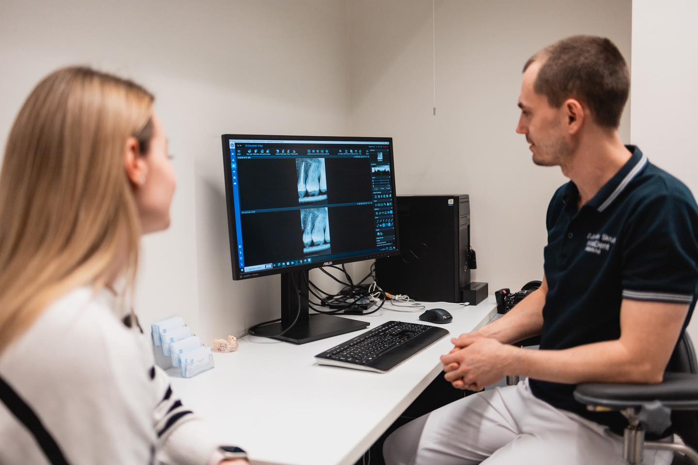
First, the doctor will ask the patient if and how the tooth has been painful, and what symptoms the patient has. The root canal specialist will then conduct an oral examination, take an X-ray, and, if necessary, perform additional tests such as sensitivity to temperature and tenderness upon biting. Once the diagnosis is confirmed, root canal treatment will begin. If the doctor has any doubts, they may decide to monitor the situation/tooth. This is often done at the very beginning of the infection, as in some cases, it can be difficult to identify exactly which tooth is causing the pain, especially when several adjacent teeth need treatment.
Stages of Root Canal Treatment:
- The doctor administers anesthesia to the patient.
- The tooth is isolated from the mouth using a rubber dam to ensure cleanliness in the treatment area.
- The tooth cavity (pulp chamber) is opened.
- The pulp chamber and root canal system are cleaned of decayed tissue and infection.
- During the procedure, additional X-rays may be taken if necessary.
- The root canal system is filled.
- The tooth is crowned or restored in another way.
Process
1
Consultation and diagnosis
The root canal dentist will conduct an initial examination, discuss the patient's complaints, assess the condition of the tooth, and, if necessary, take an X-ray for an accurate diagnosis and treatment plan.
2
Preparation for the procedure
The dentist ensures the sterility of the procedure, administers local anesthesia, and opens the tooth cavity to access the root canals.
3
Performing the procedure
The root canals are cleaned, disinfected, and filled, after which the tooth is restored with a filling or crown, depending on the patient's needs.
Pricelist
Root canal treatment prices: Check out the basic prices and book an appointment today!
All prices| SERVICE | PRICE |
| Consultation | from 60€ |
| Incisor/canine primary root canal treatment ca | 600€ |
| Incisor/canine retreatment ca | 680€ |
| Premolar root canal initial treatment ca | 700€ |
| Premolar root canal retreatment ca | 790€ |
| Molar root canal initial treatment ca | 880€ |
| Molar root canal retreatment ca | 990€ |
| The cost of restoring a tooth with a filling will be added. |
Satisfied customers
47000+

Customer reviews
First-class service, truly impressive. I will definitely come back again!

Aleksandr Shmanjov
Dr. Alina Ismailova is very pleasant and professional.

Angelica Tahker
A very polite, professional, and attentive dentist, Dr. Rasmus Peeker!
My previous visits to the dentist were quite painful, especially the removal of a wisdom tooth. However, I went to Dr. Rasmus Peeker, and the wisdom tooth extraction was absolutely painless! Highly recommend!

Nadezda Zdanova
Great dentists who do their job well, both in treatment and consultation. In particular, Dr. Ermel's advice helped me take better care of my teeth, so I don't need to visit the dentist as often as I used to.

Aleksis Anijärv
Everyone welcomed me very warmly. The doctor has always worked quickly, painlessly, and with high quality. Now I visit Hanzadent twice a year for regular check-ups to make sure everything is fine and to feel more confident!

Stiven Lipetski
Contraindications
We recommend postponing root canal treatment in the following cases, if possible: acute infections or inflammations in the mouth, serious systemic diseases, allergies to medications or materials used, insufficient oral hygiene, and extensive damage to the tooth structure or root that makes restoration impossible.
- Acute infections or inflammations in the mouth
- Serious systemic diseases (consultation with the patient’s doctor is necessary)
- Allergies to medications or materials used
- Insufficient oral hygiene
- Extensive damage to the tooth structure or root

Dentists

Dentist’s Advice
What happens if root canal treatment is not performed?
- Spread of infection – Untreated root infection can spread to the jawbone, gums, or even other parts of the body, leading to serious health issues.
- Persistent pain – Tooth pain will worsen over time and disrupt daily life.
- Tooth loss – If treatment is delayed, the tooth may become irreparable, and extraction may become the only solution.
- More expensive treatment in the future – Treating complications later may be much more complex and costly than performing root canal treatment in a timely manner.
- Body-wide complications – Oral infections can spread to the bloodstream, causing heart problems, weakened immunity, or other serious conditions.
Don’t delay! Timely root canal treatment helps avoid serious consequences and preserves the health of your teeth.
Good to know
FAQ

Mihkel 32 y.
18.12.2024
Duration of root canal treatment?
Mihkel 32 y. 18.12.2024
Answer: Sometimes the procedure can be done in one visit, sometimes it is necessary to visit several times. The endodontist may give an estimate of the duration of the treatment at the beginning of the treatment. It should be noted that the estimate may not always be 100% accurate, it is only the doctor’s guess based on his previous experience.

Laura 27 y.
18.12.2024
Pain after root canal treatment?
Laura 27 y. 18.12.2024
Answer: Post-procedure pain may occur in some cases.

Katrin 45 y.
18.12.2024
Is root canal treatment for a front tooth more complicated than for a chewing tooth?
Katrin 45 y. 18.12.2024
Answer: Incisors, canines, and premolars have only one root canal. Since the anatomy is simpler, the procedure is also a little easier. If the tooth has already been treated and retreatment is needed, this makes the procedure more complicated.

Andres 38 y.
18.12.2024
What is the cost of root canal treatment?
Andres 38 y. 18.12.2024
Answer: The cost depends on whether it is a primary root canal treatment or a retreatment of a previously treated tooth. In the first case, the treatment is more affordable. The number of root canals is also important; teeth with fewer root canals are more affordable.

Kristiina 29 y.
18.12.2024
Should I choose root canal treatment with a microscope?
Kristiina 29 y. 18.12.2024
Answer: A microscope allows the doctor to better see the small structures inside the tooth and thus ensures a better treatment outcome.

Peeter 51 y.
18.12.2024
Will my tooth heal?
Peeter 51 y. 18.12.2024
Answer: This procedure, like any other treatment, does not guarantee a cure for the problem/disease. At the beginning of treatment, the doctor can predict the outcome, whether it will be favorable or poor. If the procedure did not give the desired result, in many cases it is possible to perform a root canal operation.

15.01.2025
15.01.2025

20.02.2026
20.02.2026


